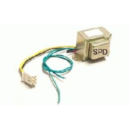
Grasslin Uni 45 Manual Transfer
The development of chemical exchange saturation transfer (CEST) and magnetization transfer (MT) contrast in MRI have enabled the enhanced detection of metabolites and biomarkers in vivo. In brain MRI, the separation between CEST and MT contrast has been particularly difficult due to overlaps in the frequency responses of the contrast mechanisms. We demonstrate here that MT and CEST contrast can be separated in the brain by the so-called uniform-MT (uMT) technique, thus opening the door to addressing long-standing ambiguities in this field. These methods could be useful for keeping track of important endogenous metabolites and for providing an improved understanding of neurological and neurodegenerative disorders.
Examples are shown from white and gray matter regions in healthy volunteers and patients with multiple sclerosis, which demonstrated that the MT effects in the brain were asymmetric and that the uMT method could make them uniform. Introduction Magnetic resonance imaging (MRI) offers a number of contrast mechanisms to noninvasively visualize the anatomical structures, physiological conditions, and functional activities of the human body. Saturation transfer (ST) provides a family of powerful and flexible contrast mechanisms, including magnetization transfer (MT) () and chemical exchange saturation transfer (CEST) (van Zijl and Yadav, 1022;;; ), to probe biomarkers, physiologically active molecules, and macromolecules in tissues and organs. Since the ST family shares a common practical procedure, in which off-resonance pre-saturation irradiation modulates the MRI signal (), those contrast mechanisms often interfere with one another, while their differentiation is highly important. For example, CEST contrast is usually produced when the pre-saturation irradiation is applied around a specific frequency offset, while MT contrast can be achieved over a broader range of frequency offsets.
Download new bodyworks 60 crack 2016 download and software. MT is also known to exhibit asymmetries with respect to the water resonance, which often prevents a conventional symmetry analysis from disentagling it from CEST contrast. Recently, it has been demonstrated that certain MT effects can be made uniform and that it is possible to separate such MT effects from the estimation of CEST effects (; ). This so-called uniform-MT (uMT) strategy is based on the finding that the uniform and efficient saturation of a strongly coupled proton spin pool can be achieved, regardless of the frequency offsets of the off-resonance pre-saturation irradiation, by irradiating the pool simultaneously at more than one frequency position (). In the brain, it has been well known that white matter and gray matter provide considerable MT effects (;; ), and MT contrast has become a routine technique, especially for the characterization of white matter diseases, such as multiple sclerosis (MS) (;;; ). Recently, several endogenous CEST contrast mechanisms have been established in the brain, which can be useful for detecting metabolites such as myo-inositol (), creatine (), and glutamate (), and accessing pH values through the so-called amide proton transfer (APT) mechanism (). Such methods have the potential for diagnosing and monitoring neurological and psychiatric disorders. On the other hand, there have so far been no conclusive studies that could quantify the interferences between the MT and CEST contrast mechanisms, although considerable uncertainties have frequently been reported in CEST measurements, including ‘negative’ CEST (; ).